
Histological Zones of Enamel Caries- (Easy Diagram Guide for Dental Students)
Understanding the 4 Histological Zones of Enamel Caries (with Diagram)
🔺️The progression of enamel caries is not random—it follows a clear histological pattern that reflects the mineral changes occurring beneath the enamel surface.
🔺️These zones are invisible clinically, but histologically they reveal the entire story of demineralization.
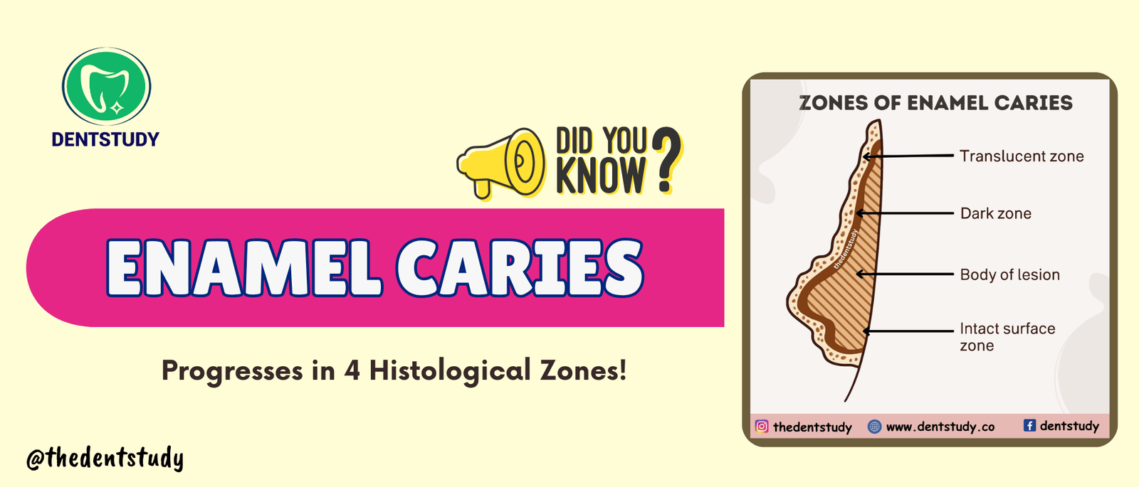
👉 The uploaded diagram illustrates the classic four zones of enamel caries:
🔹️Translucent Caries
🔹️Dark Zone
🔹️Body of Lesion
🔹️Intact Surface Zone
Would you like to continue reading?
Log in or subscribe to unlock the full premium note.
Log in to continue